
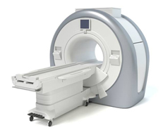
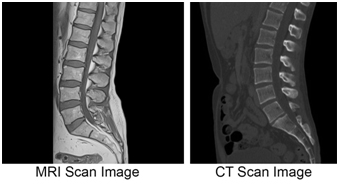
 Diagnostic Centre has 1.5 Magnetic Resonance Imaging(MRI) which is a medical imaging technique used in radiology to form pictures of the anatomy and the physiological processes of the body in both health and disease. MRI does not involve X-rays and the use of ionizing radiation, which distinguishes it from CT or CAT scans. Magnetic resonance imaging is a medical application of nuclear magnetic resonance (NMR).
MRI is the investigation of choice in the preoperative staging of rectal and prostate cancer and, has a role in the diagnosis, staging, and follow-up of other tumors.
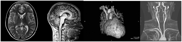
 MRI is the investigative tool of choice for neurological cancers, as it has better resolution than CT and offers better visualization of the posterior fossa. The contrast provided between grey and white matter makes MRI the best choice for many conditions of the central nervous system, including demyelinating diseases, dementia, cerebrovascular disease, infectious diseases, and epilepsy. Since many images are taken milliseconds apart, it shows how the brain responds to different stimuli, enabling researchers to study both the functional and structural brain abnormalities in psychological disorders. MRI also is used in guided stereotactic surgery and radiosurgery for treatment of intracranial tumors, arteriovenous malformations, and other surgically treatable conditions using a device known as the N-localizer.
 Cardiac MRI is complementary to other imaging techniques, such as echocardiography, cardiac CT, and nuclear medicine. Its applications include assessment of myocardial ischemia and viability, cardiomyopathies, myocarditis, iron overload, vascular diseases, and congenital heart disease.
Applications in the musculoskeletal system include spinal imaging, assessment of joint disease, and soft tissue tumors.
Hepatobiliary MR is used to detect and characterize lesions of the liver, pancreas, and bile ducts.
Magnetic resonance angiography (MRA) generates pictures of the arteries to evaluate them for stenosis (abnormal narrowing) or aneurysms (vessel wall dilatations, at risk of rupture). MRA is often used to evaluate the arteries of the neck and brain, the thoracic and abdominal aorta, the renal arteries, and the legs (called a "run-off").
![]()
Vijay Chowk, 400 meters towards Alinagar ,150 Dilezakpur, Gorakhpur, Uttar Pradesh 273001.
100 meters ahead of Chatra Sangh Chauraha, Cantt police station road, Gorakhpur.
+91-8565828541, 9335448113
